II.A Examples of Porous Media
Most materials are porous when viewed at an appropriate length scale. Examples range from porous silicon which is porous on the subnanometer scale to limestone caves and underground river systems on the kilometer scale. An anthology of examples illustrates the variability of length scales and microstructures found in porous media.
Highly porous electrochemically etched silicon has recently found much interest because of its visible luminescence which is attributed to its porous microstructure [12, 13, 50, 51, 52, 53, 54]. Figure 2 shows transmission electron micrograph of the microstructure of porous silicon [13]. Other example for microporous solids are zeolites.
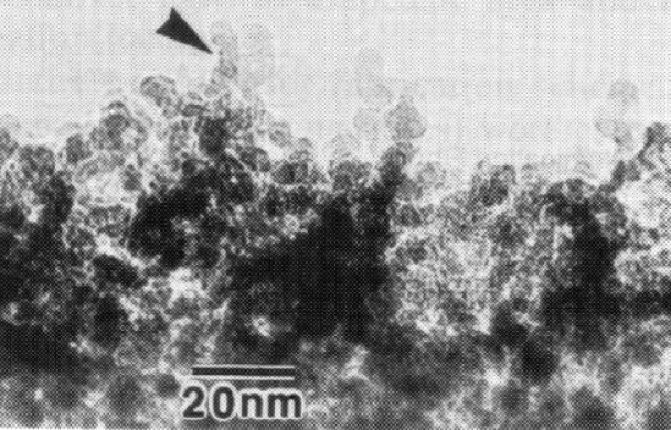
Figure 3 shows a scanning electron micrograph of a critical point dried gel consisting of ultrahigh molecular weight polyethylene. [55]
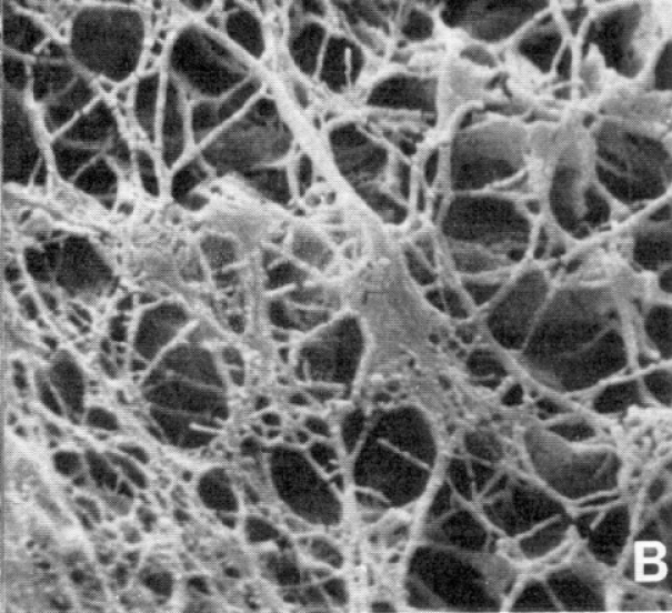
The gel was obtained from 2% solutions in decalin under agitated conditions prior to gelation, and the fibrillar structure of the polyethylene crystals reflects flow prior to gelation. Thermoreversible gels consist of a macroscopic mechanically coherent network of macromolecules that is formed and stabilized through interconnected crystals. These materials are of considerable importance in the processing of high performance polymers with very high tensile strength [55].
Figure 4 displays the microstructure of silicon
nitride ceramics consisting of elongated ![]() grains
embedded in a matrix of finer grains and a grain
boundary phase. Silicon nitride ceramics with high strength
have a fine grained elongated microstructure while materials
with a high fracture toughness are more coarse grained.
[15]
grains
embedded in a matrix of finer grains and a grain
boundary phase. Silicon nitride ceramics with high strength
have a fine grained elongated microstructure while materials
with a high fracture toughness are more coarse grained.
[15]
Wood is a strongly anisotropic natural porous medium exhibiting cylindrical pores.
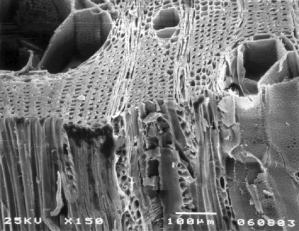
Figure 5 shows a scanning electron micrograph of a partially
cut and fractured surface from malaysian Nemesu wood which gives
an impression of the irregularities within the material.
The image shows tracheid cells of about 25![]() m in diameter
and vessel elements which are roughly an order of magnitude
larger.
m in diameter
and vessel elements which are roughly an order of magnitude
larger.
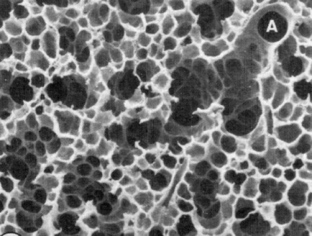
The large surface to volume ratio which is characteristic for porous media (see section 3.1 below) is essential for the function of the lung consisting of some 300 million small air chambers. Figure 6 shows the foam like structure formed by the respiratory air chambers in the lung.
As emphasized by Weibel biological porous media should not be viewed as random [56]. The general difficulty of modeling porous media as random or disordered media is further discussed in section II.B.2 below.
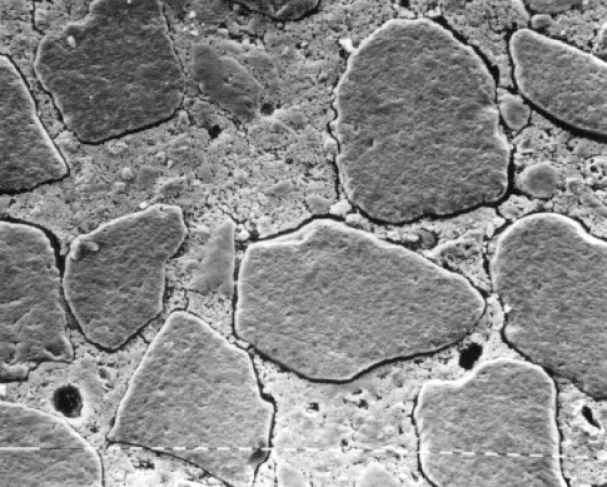
Building and construction materials such as cements and concrete
are porous media. Figure 7
shows a scanning electron micrograph of cement and lime mortar.
Each dash in the dashed line visible in the image corresponds
to ![]() m. The micrograph shows an extended network of
fissures whose properties govern the moisture transfer
and sorption properties of such materials [57].
m. The micrograph shows an extended network of
fissures whose properties govern the moisture transfer
and sorption properties of such materials [57].
An understanding of transport and relaxation in rocks, soils and other geologically important heterogeneous media is of crucial importance in hydrology, exploration geology, petroleum engineering and environmental research.
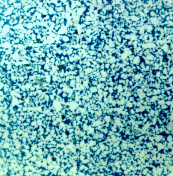
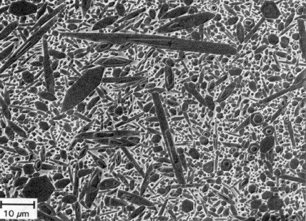
Figure 8 shows a thin section of a clastic sandstone formed by fluvial deposits. Sandstones and other sedimentary rocks have attracted much research interest, and are among the best investigated examples of porous media.
The variety of two component porous media is enormous. Most of the discussion below will focus on irregular, random media. Regular and ordered microstructures (such as in zeolites) may be considered as a special case of irregular porous media.
